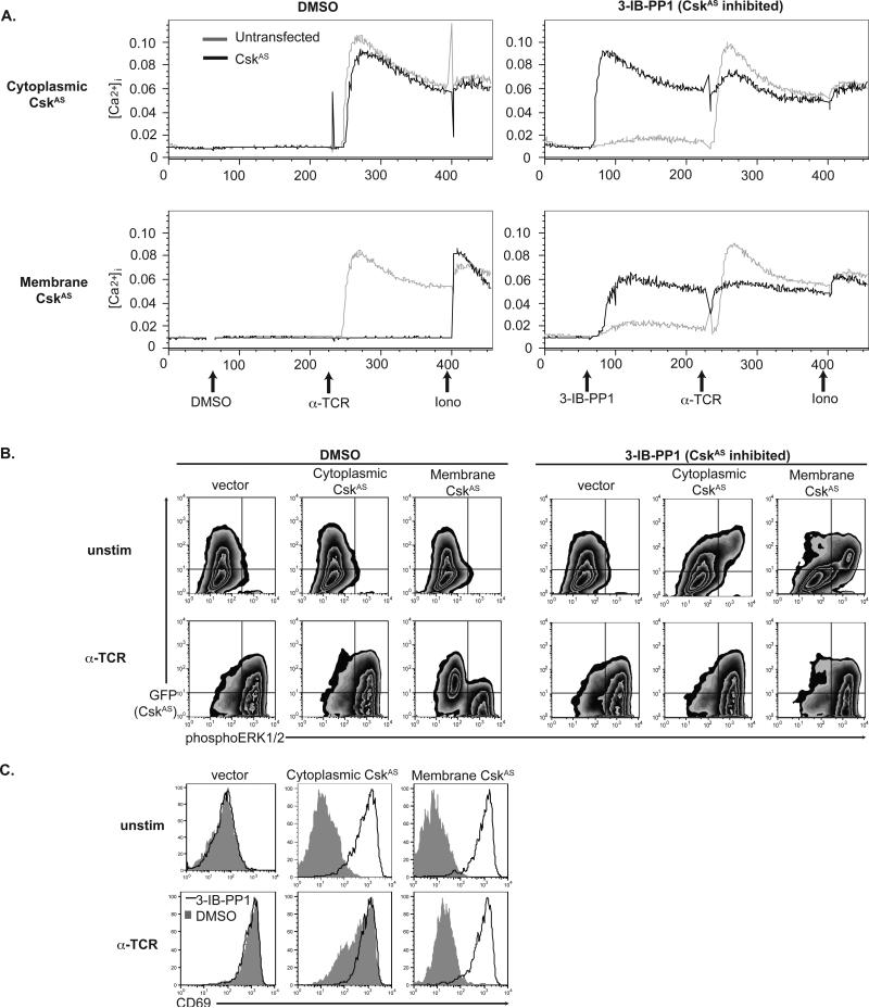
Figure 4. Distal TCR signaling and cell activation induced by CskAS inhibition.
(A) Calcium release is triggered by CskAS inhibition alone in T cells expressing cytoplasmic- (top) or membrane- CskAS (bottom). Jurkat T cells transiently cotransfected with CskAS and CD16 constructs. The ratios between Fluo-3 and FuraRed is shown for CD16− untransfected cells or for CD16+ CskAS transfected cells in response to DMSO, 3-IB-PP1, anti-TCR and ionomycin. B) Jurkat T cells expressing membrane-CskAS have impaired ERK phosphorylation that is overcome by inhibition of CskAS alone. Control or CskAS cells were serum-starved, pretreated with DMSO or 3-IB-PP1 for 15 minutes. Cells were then harvested directly (unstim) or after 2 minutes of TCR stimulation. Plots show total live cells, with GFP− untransfected cells in the bottom quadrants and GFP+ CskAS–transfected cells in the upper quadrants. (C) Upregulation of CD69 is impaired in TCR-stimulated membrane-CskAS cells, and is induced in response to CskAS inhibition alone. Transiently transfected Jurkat T cells were treated with either 3-IB-PP1 or DMSO, and were TCR stimulated for 18 hours prior to surface staining for CD69. Data are representative of two (A) or three (B, C) independent experiments.

