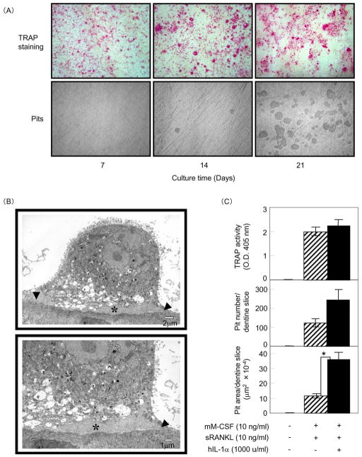
Figure 2. Osteoclast differentiation of 4B12 cells on dentine slices.
(A) 4B12 cells (5 × 10³) were cultured in the presence of mM-CSF (10 ng/ml) plus sRANKL (10 ng/ml) plus hIL-1α (1000 u/ml) on dentine slices in 24-well culture plates. The culture medium was exchanged for new culture medium every 7 days. After 7, 14, and 21 days of culture, 4B12 cells on dentine slices were stained for TRAP. After removing the 4B12 cells on dentine slices, resorption pits on the dentine surface were observed by reflective light microscopy. (B) Ultrastructural features of bone-resorbing cells formed from 4B12 cells in the presence of mM-CSF (10 ng/ml) plus sRANKL (10 ng/ml) plus hIL-1α (1000 u/ml). The cells exhibit a clear zone (arrowhead) and ruffled borders (asterisk). (C) 4B12 cells (5 × 10³) were cultured with or without hIL-1α (1000 u/ml) in the presence of mM-CSF (10 ng/ml) and sRANKL (10 ng/ml) on dentine slices for 21 days. TRAP activity and the number and total area of the resorption pits formed on the dentine slices were measured. *P<0.05 versus the culture without hIL-1α. Similar results were obtained in two independent experiments.

