
Figure 3. Time Dependent GILZ Induction by Dex in Cultured Cardiomyocytes.
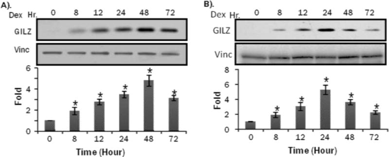
Primary cultured neonatal rat cardiomyocytes (A) or H9c2(2-1) cells (B) were treated with 1 M Dex for indicated time. Cells were harvested for Western blot to measure GILZ protein levels with vinculin (vinc) as loading control. The intensities of the bands were quantified and significant difference from 0 hr, as indicated by * (p<0.05), was determined as described in Figure 2.