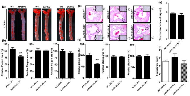
Fig. 2. Plaque area was reduced in the MARKO-LDLR−/− mice compared to the WT-LDLR−/− control mice, but not reduced in the EARKO-LDLR−/− and SARKO-LDLR−/− mice.
a. Oil-red-O staining of aortas obtained from the MARKO-LDLR−/−, EARKO-LDLR−/−, and SARKO-LDLR−/− mice and their WT-LDLR−/− littermate control mice. Aorta were stained with Oil-red-O and positive red stained area indicates plaques formed. b. Quantitation results of positive plaque area over total area from a. c. H & E staining of aortic tissues of MARKO-LDLR−/−, EARKO-LDLR−/−, and SARKO-LDLR−/− mice and their WT-LDLR−/− littermate control mice. Magnification, 100x (inset, 400x). Two arrowheads indicate the plaque area. d. Quantitation of plaque area over total area from c. e. Testosterone levels were determined in MARKO-LDLR−/− mice and their WT-LDLR−/− littermate controls. f. Testosterone levels in WT-LDLR−/−, SARKO-LDLR−/−, and EARKO-LDLR−/− mice. **p<0.01

