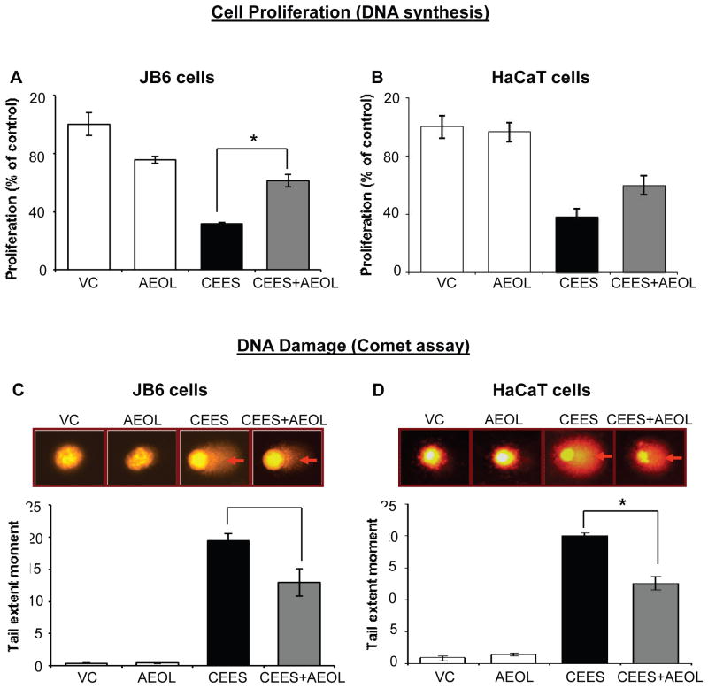
Figure 2. AEOL 10150 treatment ameliorates CEES-induced decrease in DNA synthesis and DNA damage in mouse skin epidermal JB6 cells and human skin epidermal HaCaT cells.
Mouse epidermal JB6 (A) and human epidermal HaCaT cells (B) were seeded and grown overnight in 96 well plates. These cells were then exposed to either DMSO alone (VC), 50 μM AEOL 10150 alone (AEOL), 0.25 mM CEES in DMSO (CEES), or to 50 μM AEOL 10150 1 h after 0.25 mM CEES exposure (CEES+AEOL) for 48 h. Thereafter, cells were incubated with BrdU for 2 h, fixed and DNA denatured and labeled with anti-BrdU mouse monoclonal Ab-Fab, and product was quantified by measuring the absorbance as detailed under Materials and Methods (A and B). After desired exposure and treatments as above for 2 h, DNA damage in the cells was measured via alkaline comet assay as detailed under Materials and Methods (C and D). Representative pictures show damaged DNA seen in the form of comets behind the JB6 (C) and HaCaT (D) cells, which was scored as the tail extent moment (TEM; product of tail length and percentage tail DNA). Data shown are mean ± SEM of 3-4 independent samples. *, p<0.05 as compared to CEES exposed group.

