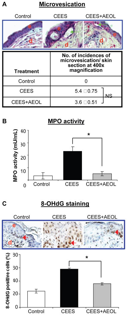
Figure 7. AEOL 10150 treatment effects on CEES-induced microvesication (A), myeloperoxidase activity (B) and DNA oxidation (C) in SKH-1 hairless mouse skin.
Mouse dorsal skin was exposed topically to either 200 μl acetone (VC) or 4 mg CEES (CEES), or treated with AEOL 10150 (subcutaneous + topical; CEES+AEOL) 1 h after CEES exposure as detailed under Materials and Methods. After 12 h of the indicated exposure or treatments, mice were sacrificed and dorsal skin tissue samples were collected and either frozen or fixed as detailed under Materials and Methods. Skin tissue sections (5 μM) from fixed tissue were processed for H&E staining and analyzed for microvesication. (A) Representative H&E stained skin sections show microvesication as well as quantified data for incidences of microvesiction (epidermal-dermal separation.. (B) Frozen skin tissue was used to determine MPO activity using a fluorescent kit from Cell Technology as detailed under Materials and Methods. (C) Skin sections (5 μm) from fixed tissue after processing were subjected to IHC for 8-OHdG as detailed under the Materials and Methods. The brown colored DAB positive nuclei were 8-OHdG positive (as shown in the representative pictures; C) and were counted in 10 randomly selected fields (400X magnification). Data presented are mean ± SEM (n=5); *, p<0.05 as compared to CEES exposed group; NS, not significant as compared to CEES group; e, epidermis; d, dermis; green arrows, epidermal-dermal separation; red arrows, 8-OHdG positive cells.

