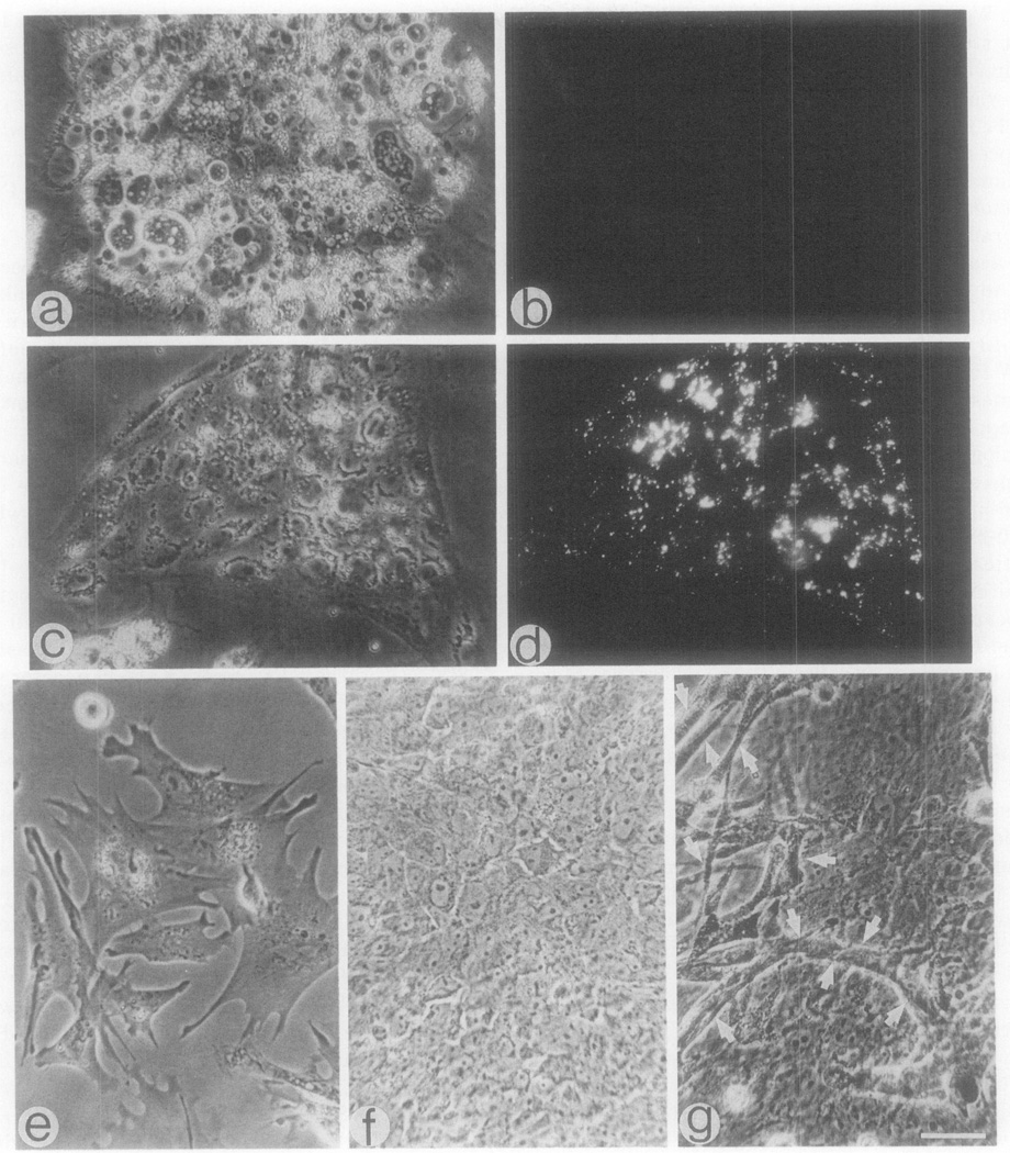
Fig. 4.
Representative cells detected in cultures of 22-hr-old chick embryos. (a–d) A 12-hr-old culture consisting of large cells which do not incorporate DiI-Ac-LDL (a, b) and islands of cells which are DiI-positive (c, d); (e–g) representative cells detected in a 7-day-old culture in addition to the cells in a and b. Bar – 30 µm.

