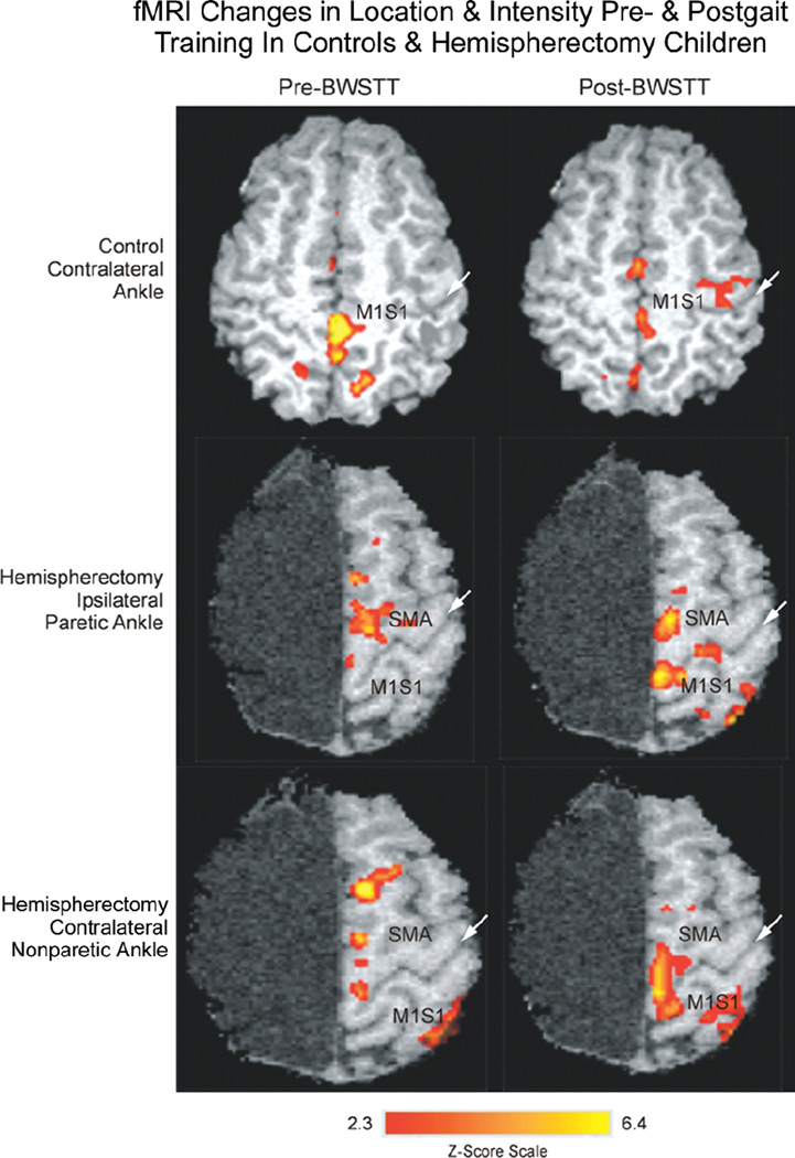
Figure 3.
Representative functional magnetic resonance imaging (fMRI) activation scans for a control (first row; 12-year-old) and posthemispherectomy patient (second and third rows; 13-year-old; right hemispherectomy for Rasmussen syndrome 8 years ear-lier) before (left column) and after (right column) body weight–supported treadmill training (BWSTT). White arrow in each panel indicates the central sulcus. Top: in a control patient, voluntary contralateral ankle movement resulted in activation of the M1S1 cortex pre-BWSTT that decreased in area and intensity post-BWSTT. Middle and lower: by contrast, in a hemispherectomy child, voluntary ipsilateral paretic and contralateral nonparetic ankle movement before gait training showed some activation of the sup-plementary motor area (SMA) and M1S1 cortex that increased in area and intensity posttherapy. Z score scale for Figures 4 and 5 is indicated at the bottom of the figure.

