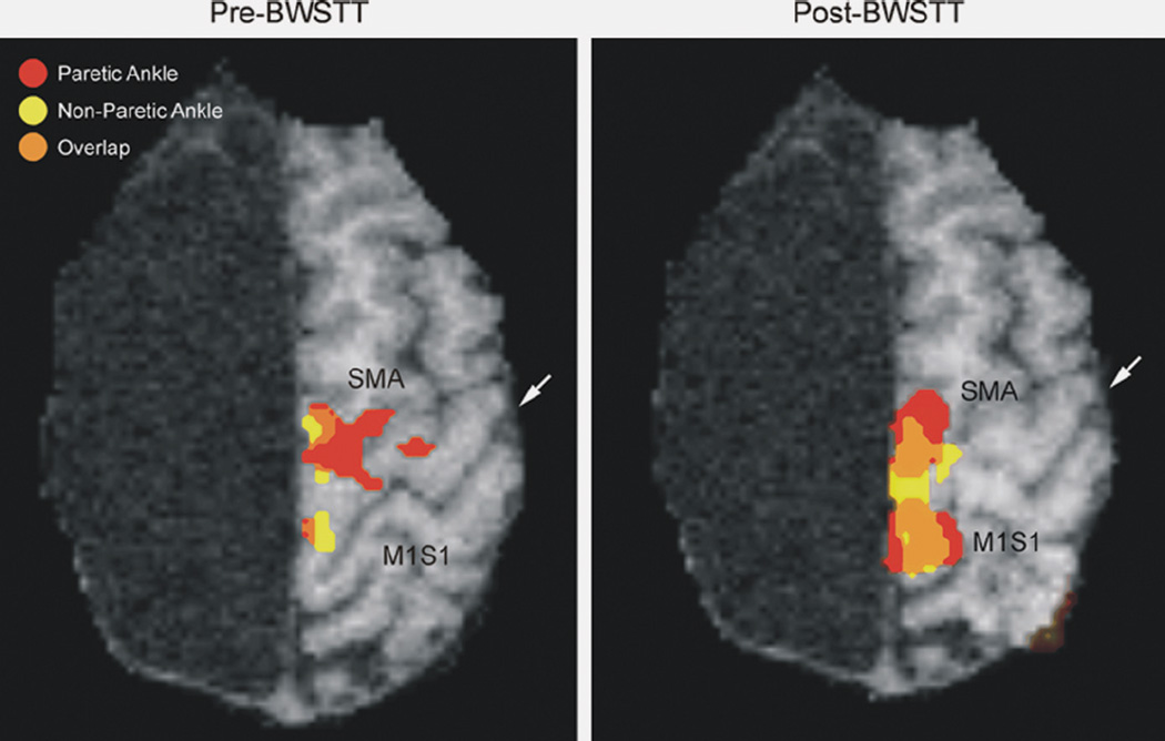
Figure 4.
Functional magnetic resonance imaging (fMRI) activation of the ipsilateral paretic ankle (red), contralateral nonparetic ankle (yellow), and areas of overlap (orange) within the supplementary motor area (SMA) and M1S1 cortex with voluntary movement in a hemispherectomy child. Pretraining, only a small area of overlap was present mostly in the SMA region (left). Posttherapy, the areas of activation increased, as did the areas of overlap, in both the SMA and M1S1 cortex. White arrow indicates the central sulcus.

