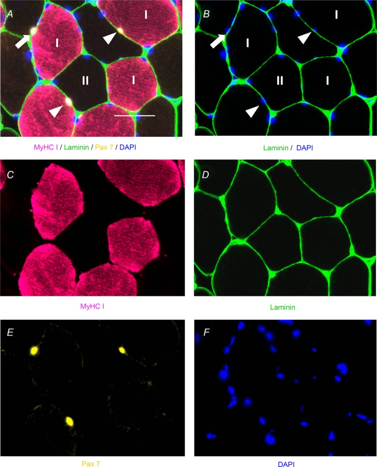
Figure 6. Representative immunohistochemical image of fibre type-specific satellite cell identification.

A, fused image demonstrating laminin (green), Pax7 (yellow), MyHC type I (pink) and 4′,6-diamidino-2-phenylindole (DAPI; blue) with MyHC type I satellite cells denoted by white arrowheads and a MyHC type II satellite cell denoted by a white arrow. Scale bar represents 50 μm. B, fused image demonstrating satellite cell location within laminin (green), costained with DAPI (blue), and MyHC type I satellite cells denoted by white arrowheads and a MyHC type II satellite cell denoted by a white arrow. C, single-channel image demonstrating MyHC type I (pink). D, single-channel image demonstrating laminin (green). E, single-channel image demonstrating Pax7 (yellow). F, single-channel image demonstrating DAPI (blue).
