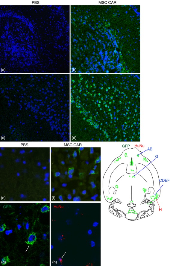
Figure 2.

Brain localization of central nervous system (CNS) -targeted mesenchymal stromal cells (MSC)s in naive mice following intranasal delivery. Human MSCs were instilled via a unilateral intransal (i.n.) administration and the distribution of green fluorescent protein (GFP) or human nuclei (HuNu) immunofluorescence in horizontal cryosections of the brain of naive mice was studied 24 hr after delivery. The schematic depicts a selective GFP and HuNu immunofluorescence (indicated by green and red spots) in various brain regions. Confocal microscopy reveals that GFP immunofluorescence (green) is present in the internal plexiform layer of the olfactory bulb (b) ectorhinal cortex (d) and Purkinje cells in the cerebellum (f) in MSC CARαMOG-treated naive mice. The corresponding areas in PBS-treated naive mice are (a) ,(c,) and (e), respectively. Cell nuclei (blue) are stained with DAPI. Original magnifications 10× (a–f) and 40× (g,h). Immunofluorescence microscopy reveals that GFP fluorescence (green) and HuNu fluorescence (red) are both present in the ectorhinal cortex.
