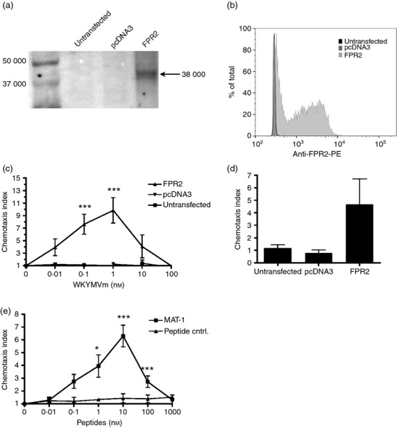
Figure 5.

The peptide MAT-1 induces chemotaxis using the formyl peptide receptor 2 (FPR2) receptor. (a) Western blot using anti-FPR2 antibody with different HEK 293 cells shows FPR2 as a 38 000 molecular weight (MW) band. (b) Cytometric analysis of FPR2 expression confirms expression in FPR2-transfected cells. (c) Chemotaxis was assessed using increasing concentrations of WKYMVm in FPR2-transfected, pcDNA3-transfected, and untransfected HEK 293 cells. Chemotaxis indices at each WKYMVm concentration with FPR2-expressing cells were compared with a corresponding concentration with pcDNA3 and untransfected cells. (d) Chemotaxis was assessed using 10 nm MAT-1 with FPR2-transfected, pcDNA3-transfected, and untransfected HEK 293 cells to confirm specificity for FPR2. (e) Chemotaxis assays with FPR2-transfected cells compared MAT-1 with the negative peptide control at the indicated concentrations. Error bars represent SEM (n = 3, *P < 0·05, **P < 0·01, ***P < 0·001).
