
Figure 5.
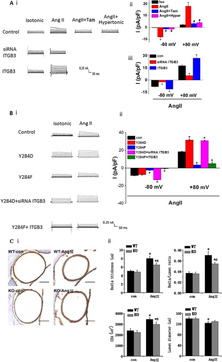
Involvement of integrin β3 in ICl.AngII and ClC-3 knockout attenuates cerebrovascular remodelling in Ang II-infused hypertensive mice. (A) In BASMCs, Ang II (200 nM) induced an outward rectifying current Cl− current (ICl.AngII) in isotonic solution, which was completely inhibited in hypertonic solution (Hyper) and by tamoxifen (Tam, 10 μm), indicating that this current was a volume-regulated Cl− current. ICl.AngII was also completely blocked by integrin β3 siRNA (siRNA ITGB3) and significantly enhanced by integrin β3 cDNA(ITGB3). (i) Representative traces of ICl.AngII. (ii) Mean current densities of control group with different treatment measured at −80 mV (downward bars) and +80 mV (upward bars). (iii) Mean current densities of angiotensin II-treated control, siRNA ITGB3 and ITGB3 groups at −80 mV (downward bars) and +80 Mv (upward bars; *P < 0.05 vs. control group, # P < 0.05 vs. Ang II group, n = 5). (B) ICl.AngII was regulated by Tyr284 phosphorylation in ClC-3. In ClC-3 Y284D and Y284F cells, ICl.AngII was enhanced and inhibited respectively. Integrin β3 cDNA transfection did not alter Y284F effect on ICl.AngII. Similarly, integrin β3 siRNA transfection did not reverse Y284D effect on ICl.AngII. (i) Representative traces of Cl− current. (ii) Mean current densities measured at −80 mV (downward bars) and +80 mV (upward bars) (*P < 0.05 vs. control group, n ≥ 6). (C) ClC-3 knockout (ClC-3−/−) ameliorated the morphological changes of mice cerebral basilar artery occurring during hypertension. (i) wt (ClC-3+/+) mice and ClC-3−/− mice were treated with or without Ang II. Vascular media smooth muscle was specifically stained as brown with α-actin antibody. Scale bars, 50 μm. Representative pictures are shown. (ii) Statistical analysis of media thickness, internal lumen diameter, media-to-lumen ratio and medial CSA (n = 6 per group, *P < 0.05, Ang II mice vs.control mice; #P < 0.05, wt-AngII mice vs. ClC-3−/−-AngII mice).