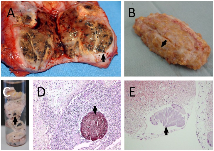
Figure 1. Different grains obtained from eumycetoma and actinomycetoma lesions.
In this figure different grain types are shown. A: An eumycetoma surgical excision with numerous black grains, indicative for M. mycetomatis. B: An actinomycetoma surgical biopsy with numerous yellow grains, indicative for S. somaliensis. C: Grains of Madurella mycetomatis fixed in formalin. D: Histological slide of a Madurella mycetomatis grain inside subcutaneous tissue. The grain is clearly seen as a round brown structure (arrow) (×100). E: Histological slide of a S. somaliensis grain inside subcutaneous tissue (arrow) (×400).

