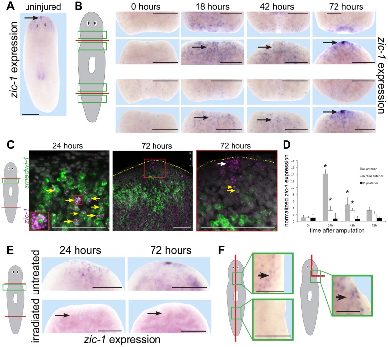
Figure 2. Injury induces expression of zic-1 in neoblasts and in the regenerating anterior pole.
(A–B) in situ hybridizations to detect expression of zic-1 in uninjured or regenerating animals as indicated. (A) zic-1 was expressed in the head region and head tip in uninjured animals (arrow). (B) zic-1 was expressed preferentially near anterior-facing versus posterior-facing amputation sites by 18 hours and is expressed at the regenerating anterior pole by 72 hours after surgery (arrows). (C) Double fluorescence in situ hybridizations (FISH) to detect co-expression of zic-1 (magenta) with smedwi-1 (green) mRNA in tissue regions near anterior-facing amputation sites. Hoechst staining shows nuclei (gray). By 24 hours after injury, numbers of zic-1+/smedwi-1+ cells increased substantially (13/29 of zic-1+ cells were smedwi-1+, n = 3 animals at 0 hours to 506/530 of zic-1+ cells were smedwi-1+, n = 4 animals at 24 hours) and by 72 hours zic-1 was expressed at the anterior pole in non-neoblast cells lacking smedwi-1 mRNA expression (10/10 cells, n = 3 animals). Arrows indicate cells co-expressing (yellow) and not singly-expressing the two genes (white). Red box indicates enlarged region of a zic-1+/smedwi-1+ cell (24 hours, inset) or anterior pole region (72 hours, right). Yellow dotted line shows anterior edge (72 hours). (D) qPCR to measure zic-1 expression versus gapdh in FACS-sorted X1 or X2/Xins cells purified as in Fig. S2A from tissue near anterior or posterior-facing injury sites as in Fig. 1B in a time series relative to expression at 5 minutes post surgery. Error bars are standard deviations. (E) Injury-induced zic-1 expression was eliminated in animals lethally irradiated with 6000 Rads immediately prior to amputation (arrows). (F) Early wound-induced expression of zic-1 occurred independently of anterior removal or midline disruption. Left, laterally amputated animals activated zic-1 expression near the injury site preferentially in animal anterior versus posterior (11/12 animals probed). Right, asymmetric anterior wedge removed tissue without midline or anterior pole disruption and resulted in expression of zic-1 by 18 hours after surgery (6/7 animals probed). Unless otherwise noted, all panels represent at least 4 of 5 animals probed. Cartoons show surgeries and enlarged regions. Anterior, top. Bars, 250 microns (A, B, E) and 75 microns (C, F).

