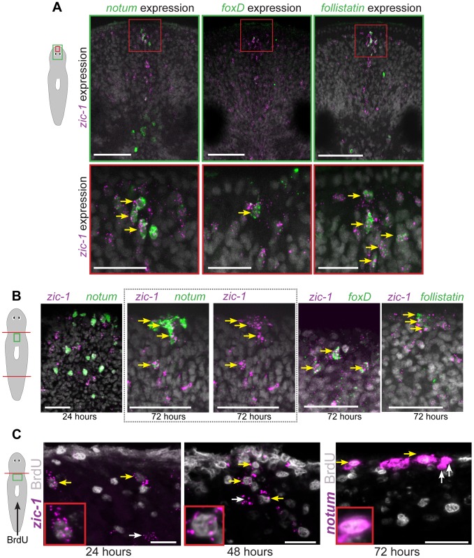
Figure 3. zic-1 is co-expressed with notum, foxD and follistatin in neoblast descendent cells at the anterior pole.
(A) Double FISH to detect expression of zic-1 (magenta) and either notum, foxD, or follistatin (green) in at the anterior pole of uninjured animals with nuclei stained using Hoechst (gray). Cartoon depicts fields of view in green (top) or red (bottom) borders. (B) Double FISH as in (A) detecting expression in regenerating fragments at the indicated times after decapitation. Dotted box, contains image of anterior pole showing zic-1 and notum staining (left) and corresponding image of only zic-1 staining (right). zic-1 is not highly co-expressed with notum at 24 hours (9/142 notum+ cells were zic-1+, n = 3 worms), but is co-expressed with notum at the anterior pole by 72 hours after surgery. At 72 hours in the regenerating anterior pole, 62.1±5.3% zic-1+ cells co-expressed notum (n = 3 worms), 91.7±11.8% zic-1+ cells co-expressed foxD (n = 5 worms), and 56.1±28.6% zic-1+ cells co-expressed follistatin (n = 6 worms). (C) Animals were injected with bromodeoxyuridine (BrdU) two days prior to decapitation, then fixed at the indicated times and probed by in situ hybridization and immunostaining to detect BrdU and either zic-1 or notum. This pulse of BrdU labeled 35.2±16.6% of zic-1+ cells at 24 hours in the parenchyma (n = 5 worms, 440 zic-1+ cells counted), and 80.4±20% of zic-1+ cells at the anterior pole by 48 hours (n = 5/6 worms with anterior poles, n = 49 zic-1+ cells counted), and labeled notum+ cells at the anterior pole by 72 hours. Yellow arrows indicate co-labeled cells, white arrows indicate single-labeled cells. Cartoons show surgeries and enlarged region. Anterior top. Bars, 20 microns (C), 30 microns(A bottom panels, B), 75 microns (A top panels).

