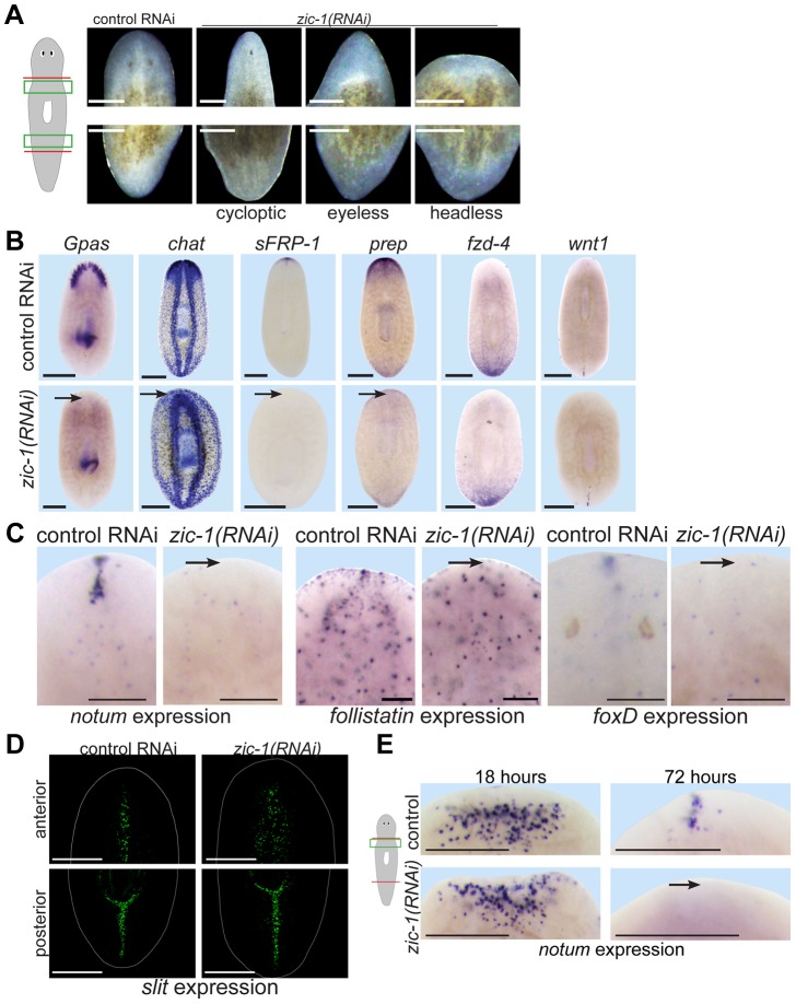
Figure 4. zic-1 is required for anterior pole formation and head regeneration.
(A) Animals 8 days after amputation of heads and tails after treatment with control dsRNA or zic-1 dsRNA, cartoon shows enlarged regions of the anterior and posterior blastemas. Inhibition of zic-1 function caused anterior regeneration defects such as cyclopia (24%, n = 74 animals scored), lack of eyes (12%, n = 74 animals scored), and head regeneration failure (63%, n = 90 animals scored). (B–D) in situ hybridizations of animals fixed 8 days after amputation. (B) The anterior region of headless zic-1(RNAi) regenerating animals lacked a brain (gpas, 2/2 animals probed; chat, 4/4 animals probed) and expression of head tip marker (sFRP-1, 11/11 animals probed) and head region marker (prep, 8/8 animals probed) and did not express posterior markers (fzd-4, 7/7 animals probed; wnt1, 7/7 animals probed). (C) zic-1(RNAi) animals failed to express notum (6/6 animals probed), follistatin (4/5 animals probed), and foxD (14/14 animals probed) at the anterior pole after 8 days of regeneration. (D) zic-1(RNAi) animals had a laterally expanded midline in their anterior but not their posterior, as assayed by slit expression (4/4 animals). (E) zic-1 is required for anterior pole formation by 72 hours and not for early wound-induced notum expression at 18 hours of regeneration. In situ hybridizations to detect notum expression after treatment with control or zic-1 dsRNA, amputation of heads and tails, and fixation at 18 or 72 hours. zic-1 inhibition prevented expression of notum at the anterior pole by 72 hours (8/8 animals) but had no effect on its expression at 18 hours (122±53 notum+ cells in control and 108±35 notum+ cells in zic-1(RNAi) animals, 8 animals probed, p = 0.59 t-test). Cartoon shows surgery and enlarged regions. Bars, 100 microns (C), 250 microns (A), 300 microns (B, D, E).

