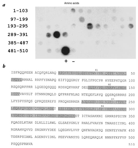
Figure 3.
Multiple IgE-binding regions identified on the Ara h 3 allergen. (a) The Ara h 3 primary sequence was synthesized as 15 amino acid–long peptides offset from each other by eight residues. These peptides were probed with a pool of serum IgE from peanut-hypersensitive patients. The position of the peptides within the Ara h 3 protein are indicated on the left. (+) indicates an immunodominant peptide from Ara h 2 that served as a positive control, and (–) indicates a peptide synthesized to serve as a negative control. (b) The amino acid sequence of the Ara h 3 protein is shown. The shaded areas (R1–R4) correspond to the IgE-binding regions shown in a.

