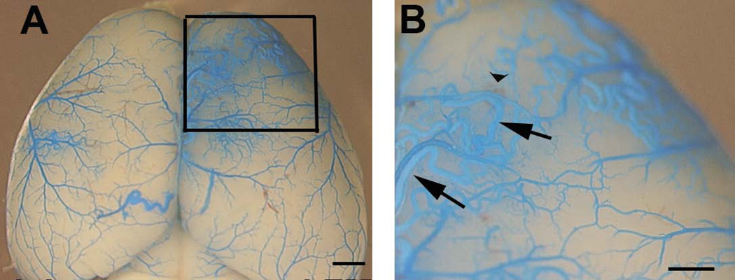
Fig. 5. Developmental onset AVMs in the postnatal brain of Eng2f/2f;SM22α-Cre mice.
(A) Representative images of latex dye casting show the AVM vessels (squared region) in the brain of 5-week-old Eng2f/2f;SM22α-Cre (B) Enlarged images of dotted boxes shown in (A). Arrows indicate latex-casted veins. Arrow head indicates hemorrhage. Due to the particle size in the latex, the dye enters the vein after intra-left cardiac perfusion only when the A–V shunts are present. Scale bars: 1 mm in (A) and 500 µm in (B).

