Figure 1.
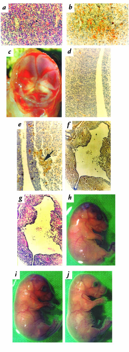
(a and b) H&E staining (a) and antifibrin(ogen) immunostaining (b) of adjacent P0.5 FVII+/–/TFPIδ/δ liver sections indicating interstitial hemorrhage and diffuse fibrin deposition. (c) A top view of the brain from a FVII+/–/TFPIδ/δ E17.5 embryo after removal of the skin and skull plate, demonstrating pools of blood on top of an opaque flattened brain. (d–f) Antifibrin(ogen) immunostaining of a E17.5 wild-type (d) or FVII+/–/TFPIδ/δ brain (e, f). Fibrin deposition (arrow) and initial brain matter degeneration is seen in e, further degeneration and fibrin deposition in f. (g) H&E staining of the corresponding section shown in f demonstrating hemorrhage. (h–j) Gross appearances of an E17.5 FVII+/+/TFPIδ/δ embryo (h) displaying growth retardation, bruising on the head, and the lack of a tail; of a E17.5 FVII+/–/TFPIδ/δ embryo (i) with less severe growth retardation and bruising; and an E17.5 FVII–/–/TFPIδ/δ embryo with normal appearance (j). E17.5, embryonic day 17.5; H&E, hematoxylin and eosin; TFPI, tissue factor pathway inhibitor.

