
Figure 2.
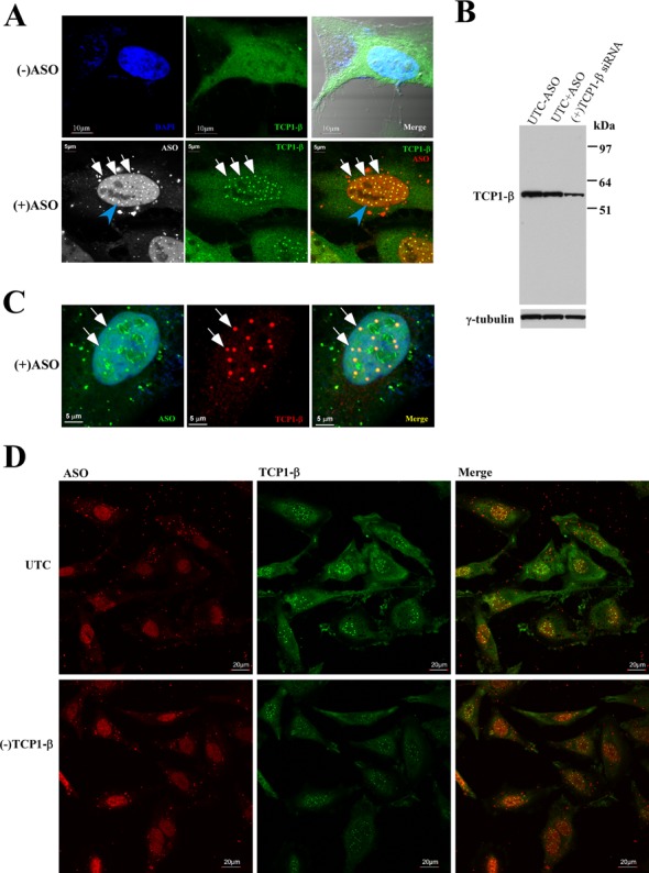
TCP1-β localizes in nuclear PS-bodies upon PS-ASO transfection. (A) Immunofluorescence staining of TCP1-β in control HeLa cells [(−)ASO] or HeLa cells transfected with 60-nM Cy3-labeled PS-ASO ISIS446654. The co-localization of TCP1-β (FITC) and PS-ASO in nuclear PS-bodies is indicated with white arrows. The scale bars: upper panels, 10 μm; lower panels, 5 μm. (B) Western analysis of TCP1-β protein in control cells transfected with (UTC+ASO) or without (UTC−ASO) 60-nM ASO ISIS116847 for 24 h, or in cells transfected with 5-nM TCP1-β siRNA for 24 h. TCP1-β protein was detected using antibody ab92756 (Abcam). γ-tubulin served as a loading control. (C) The PS-body localization of TCP1-β was not due to channel crosstalk. HeLa cells were transfected with 60 nM of FITC-labeled PS-ASO ISIS256903 and stained for TCP1-β (AF647) as described in panel (A). Scale bars, 5 μm. (D) Reduction in levels of TCP1-β did not completely block the formation of PS-bodies. HeLa cells treated with TCP1-β siRNA [(−)TCP1-β] for 24 h were transfected with 60-nM ISIS446654 for 5 h and stained for TCP1-β protein (FITC). UTC: untreated control cells. Scale bars: 20 μm.