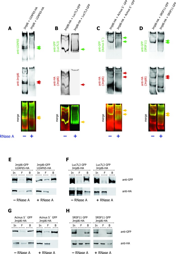
Figure 4.

Interaction of Jmjd6 and SR–proteins is RNA dependent. The bands representing Jmjd6–SR–protein complexes in native gels disappear upon RNase A treatment. Both, Jmjd6 and U2AF65 were not found migrating into the gel after RNase treatment (A). Luc7L3 entered the gel after RNase A treatment, but was not able to recruit Jmjd6 (B). In case of Acinus S′ (C) and SRSF11 (D) the position of the Jmjd6–SR–protein complex was shifted upon RNase treatment. Complexes are indicated by arrows (A–D, red and green arrows indicate single colour bands that are co-localised as seen with yellow arrows in merged images). + = RNase A treatment; − = no RNase A treatment. RNase A treatment of 293T cell lysates from cells transfected with plasmids encoding GFP-tagged and HA-tagged proteins as indicated prior to anti-GFP pulldown, SDS-PAGE and western blot resulted in a breakdown of interaction of Jmjd6 with the SR–proteins U2AF65 (E), Luc7L3 (F) and Acinus S′ (G) but not with SRSF11 (H). In = input, F = flow-through, B = beads. Upper panels show precipitation of GFP-tagged proteins, lower panels show co-precipitation of HA-tagged proteins.
