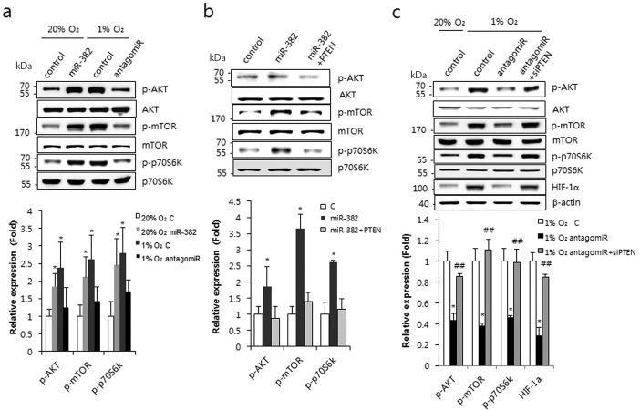
Figure 5.
miR-382 activated the AKT/mTOR signaling pathway. (a) MKN1 cells were transfected with miR-382 (50 nM) or with antagomiR-382 (70 nM) and exposed to normoxic or hypoxic conditions. Western blot analysis was performed using antibodies against the phosphorylated or native forms of AKT, mTOR and p70S6K. (b) MKN1 cells were transfected with PTEN full-length plasmid and/or miR-382 oligomers and western blot analysis was performed at 24 h after transfection. Expression of the phosphorylated or native forms of AKT, mTOR and p70S6K was determined using appropriate antibodies. (c) MKN1 cells were transfected with siRNA PTEN and/or antagomiR-382 (70 nM) and exposed to normoxic or hypoxic conditions. Western blot analysis was performed using antibodies against HIF-1α and the phosphorylated or native forms of AKT, mTOR and p70S6K. Relative protein expression (phosphorylated/unphosphorylated protein) was calculated and plotted. β-actin was used for internal control for HIF-1α in (c). Three independent experiments were performed in triplicate. *P < 0.05, **P < 0.01 or ***P < 0.001 versus 20% O2 or normoxic control. ##P < 0.01 versus hypoxic control.

