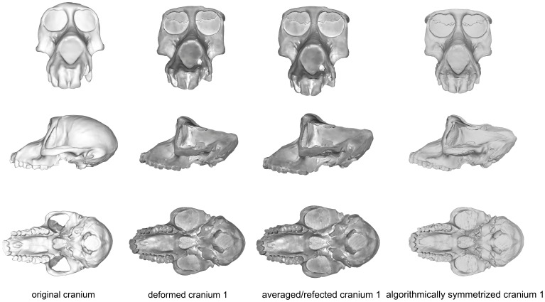
Figure 4. Comparison of the original cranium (left column), deformed cranium 1 (second column), reflected & averaged cranium 1 (third column) and algorithmically symmetrized cranium 1 (right column) in anterior (top), lateral (middle) and basal (bottom) views.
Reflected & averaged specimens do not appear perfectly symmetrical as only bilateral landmark points were used in this computation, rather than semilandmark curves or patches.

