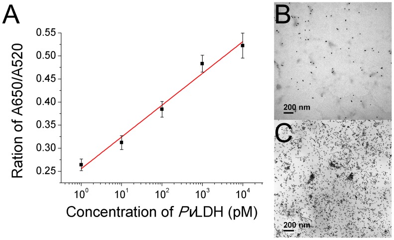
Figure 4. Detection of PvLDH in the human serum sample.
(A) The calibration curve of the sensing solutions containing varying concentrations of PvLDH in the serum sample. Points and error bars represents the means and standard deviations, respectively, of three repeated measurements. (B) The TEM image of the AuNP solution containing the serum sample. (C) The TEM image of the AuNP solution containing 10 nM of PvLDH in the serum sample.

