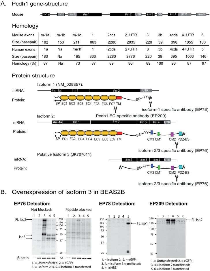
Figure 1. Pcdh1 gene-structure and isoforms.
(A) Two novel exons were detected at the 5′end of PCDH1, and one novel exon on the 3′end of Pcdh1. Mouse Pcdh1 exons share high homology with human PCDH1 exons. Corresponding isoforms (isoforms 1, 2 and putative isoform 3) and their protein structures are depicted. Only isoform 2 and putative isoform 3 contain evolutionary conserved motifs (CM1–3). Two antibodies were generated against sequences in the intracellular tail of both isoform 1 (EP78) and isoform 2 (EP76) of Pcdh1. An antibody directed against the extracellular domains of Pcdh1 (EP209) was generated previously [12]. (B) Western blot of lysates of BEAS2B cells overexpressing the open reading frame of Pcdh1 isoform 3, using antibody EP76 for detection as indicated (left hand panel) including a pre-incubation with the immunizing peptide (‘Peptide blocked’) as a control for specificity (right hand panel). Western blots using EP78 and EP209 antibodies for detection are also included as indicated. cds = coding sequence; UTR = Untranslated region; SP = Signal peptide (amino-acid (aa) 1–57); EC = Extracellular Cadherin domain; TM = Transmembrane domain; CM = Conserved Motif; PDZ-BS = PDZ-domain Binding Site; S = Serine residue; Y = Tyrosine residue; EP209 = Binding site for antibody directed against the extracellular domain of Pcdh1; EP78 = Binding site for antibody directed against a specific intracellular sequence of isoform 1, encoded by exon 2; EP76 = Binding site for antibody directed against the intracellular domain present in isoforms 2 and 3 and encoded by exon 4; FL = full length. Genbank accession numbers of novel transcripts are provided in Table 2.

