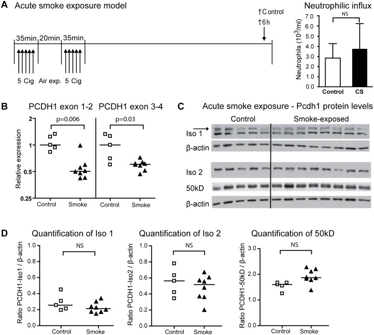
Figure 4. Pcdh1 mRNA expression levels decrease after acute smoke exposure.
(A) In the acute smoke exposure model, mice were exposed to 10 cigarettes, with a 20 min rest period in between two sessions of 5 cigarettes, or to air as a control. Low numbers of neutrophils were observed in the lung, 6 h the last exposure. (B) Pcdh1 exon 1–2 and 3–4 mRNA expression levels were determined in the cigarette smoke exposed group (closed symbols) and compared to the air-exposed group (open symbols). (C) Western blot of Pcdh1 isoform 1 (iso 1, EP78-antibody, specific band indicated with an arrow), isoform 2 (iso 2, EP76-antibody) and the 50 kD fragment (EP76-antibody) in lung homogenate of mice exposed to cigarette smoke (smoke) or air (control). (D) Densitometric quantification of Pcdh1 expression levels of full-length isoform 1 and 2, and the 50 kD fragment, relative to β-actin. Cig = cigarette; † = time-point at which mice are sacrificed; CS = cigarette smoke.

