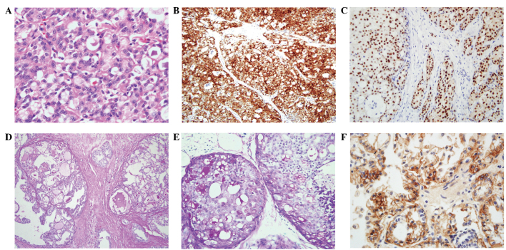
Figure 2.
Histological findings of secretory breast carcinoma. (A) Tumor cells are large and polygonal, and possess eosinophilic cytoplasm and round nuclei. Secretory material is observed in the spaces (stain, H&E; magnification, ×400). (B) S-100 protein staining is positive in the nucleus and the cytoplasm of the tumor cells (stain, S-100 antibody; magnification, ×100). (C) Secretory materials are positive for PAS (stain, PAS; magnification, ×100). (D) PAS is positive in the secretory material in ductal carcinoma in situ (stain, PAS; magnification, ×200). (E) Staining for ER is positive in in situ and invasive ductal carcinoma (stain, ER antibody; magnification, ×200). (F) Membranous staining for EGFR (stain, EGFR antibody; magnification, ×400). H&E, hematoxylin and eosin; ER, estrogen receptor; PAS, periodic acid-Schiff; EGFR, epidermal growth factor receptor.

