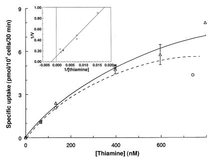
Figure 3.
Concentration dependence of high-affinity thiamine uptake in normal fibroblasts. Cells were labeled with 66 nM [3H]thiamine diluted with unlabeled thiamine for 30 min at 37°C. Values are corrected for nonspecific uptake by subtracting counts associated with the presence of 500-fold excess unlabeled compound. Results are shown for two separate experiments (solid line with triangles and dotted line with circles), done on different days with different wild-type cell lines. Inset: double reciprocal plot; the 800-nM point was eliminated as an outlier. All other data from the rectangular plot were included. The least-squares regression line gives an apparent Km of 550 nM; apparent Vmax for uptake is 11 pmol/106 cells/30 min. Each point is the mean of duplicate wells ± range (error bars).

