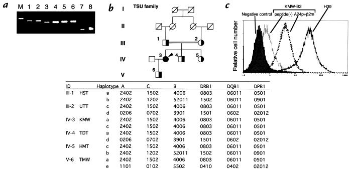
Figure 1.
(a) Expression of the mRNA of HLA-A (lanes 1 and 2), HLA-B (lanes 3 and 4), HLA-C (lanes 5 and 6), and G3PDH (lanes 7 and 8) of the lymphocytes of a healthy donor (lanes 1, 3, 5, and 7) and those of KMW (lanes 2, 4, 6, and 8). M represents a molecular weight marker. HLA-A, HLA-B, and HLA-C fragments were amplified by PCR from the cDNA derived from lymphocytes. The PCR products were sequenced by automated sequencing. (b) Consanguineous pedigree and HLA haplotypes of the TSU family. The filled circle represents an affected woman (IV-3, KMW). Partially filled circles (women) and partially filled squares (men) indicate the family members whose samples were analyzed. HLA class I and class II were typed by the sequencing-based typing system as described in Methods, and HLA haplotypes were estimated. (c) Expression of HLA class I molecules on KMW-B2 cells, a B-cell line of KMW, after incubation in the presence or absence of the A24 consensus peptide (A24p, YYEEQHPEL) and β2m. The KMW-B2 cells were preincubated in the presence or absence of 100 mg/ml A24p and 10 mg/ml human β2m for 18 h. KMW-B2 cells or H39 cells, a normal control B-cell line, were stained by FITC-conjugated mouse IgG (shaded histogram) or FITC-conjugated w6/32 and analyzed using the EPICS XL flow cytometer (Coulter Corp.). HLA, histocompatibility leukocyte antigen.

