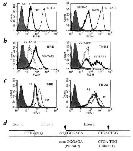
Figure 2.
BRE and TND-3 cell lines are TAP1 deficient. (a) Cells were labeled with the MAB W6/32 and analyzed by flow cytometry. Closed histograms, isotype control. Thick lines: left, BRE fibroblast cell line from patient 1; right, TND-3 EBV-B cell line from patient 2. Dotted lines: left, STF-1 TAP2-deficient fibroblast cell line; right, ST-EMO TAP2-deficient EBV-B cell line. Thin lines: left, STF-6 normal fibroblast cell line; right, ST-EAH normal EBV-B cell line. (b) Cells were infected overnight with TAP1 or TAP2 recombinant vaccinia viruses, labeled with W6/32, and analyzed by flow cytometry. Left, BRE cells from patient 1; right, TND-3 cells from patient 2. (c) BRE and TND-3 cells were incubated overnight at 26°C in culture medium (RPMI 1640 supplemented with 10% FCS; Life Technologies) in the presence of 100 μg/ml synthetic peptides, then labeled with MAB W6/32 and analyzed by flow cytometry. Closed histograms, isotype control. Thick lines, incubation without peptide. Thin lines: P1, HLA-A2402-specific peptide; P2, HLA-A2601-specific peptide. (d) Molecular genetic characterization of the mutation. Upper sequence, normal TAP1 sequence; lower sequences, corresponding TAP1 sequences in patients 1 and 2. G indicates the exon 2 nucleotide involved in the splicing reaction between exon 1 and exon 2, and arrows point to the positions of the two mutations.

