Figure 2.
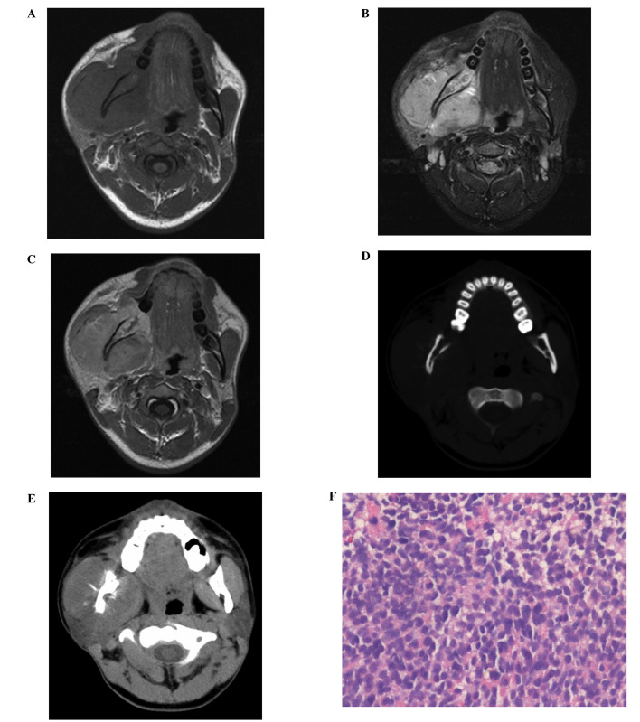
(A, B and C) Magnetic resonance imaging of the head and neck revealed a soft tissue mass arising from the right mandibular ramus, which occupied the right masseter compartment. The tumor was (A) isointense to the normal muscle on the T1WI and (B) hyperintense on the T2WI. (C) The tumor was enhanced heterogeneously following the intravenous administration of gadolinium on the contrast-enhanced T1WI. Computed tomography of the head and neck in the (D) bone and (E) soft-tissue revealed cortical destruction and a sunburst-like periosteal reaction of the right mandibular ramus.(F) A hematoxylin and eosin-stained tumor tissue section revealed small cells, which were diffuse in distribution across the majority of the view (magnification, ×10). T1WI, T1-weighted images; T1WI, T2-weighted images.

