Figure 6.
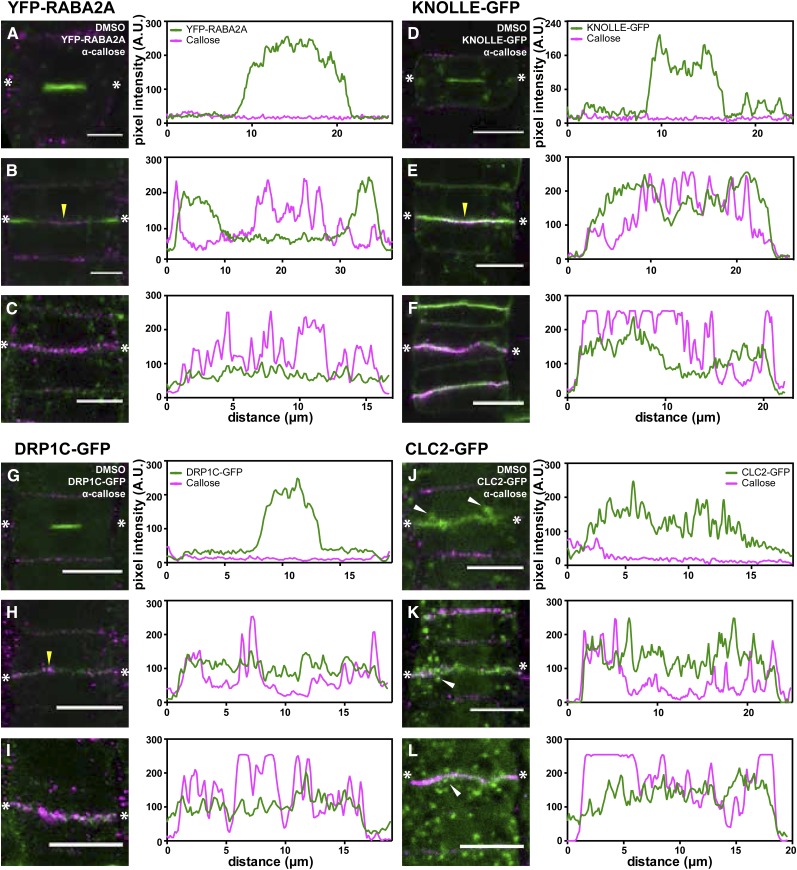
Differential vesicle accumulation occurs during callose deposition at the cell plate. Immunostaining of callose is shown with respect to the localization of vesicle markers involved in cell plate formation. Representative cells at different cell plate maturation stages are shown, and fluorescence intensities are plotted in line profiles, as indicated with the two asterisks, reflecting the fluorescence at the center of the cell plate. Throughout, α-callose is marked in magenta, while the respective fluorescent markers are indicated in green. A to C, YFP-RABA2A-labeled vesicles largely accumulate at the center of the cell during early stages, in which callose is not detectable (A). In later stages (B), YFP-RABA2A is localized at the leading edge of the cell plate, while callose started accumulating (yellow arrowhead). With the callose accumulation increasing along the cell plate, the presence of YFP-RABA2A is reduced concomitantly (C). D to F, Similar to YFP-RABA2A, KNOLLE-GFP accumulates at the center of the cell during the FVS (D). However, unlike for YFP-RABA2A, the accumulation of KNOLLE-GFP remains the same during callose deposition (E and F). G to I, The DRP1C-GFP accumulation pattern is similar to that of KNOLLE-GFP. Vesicles accumulate along the cell plate in the FVS (G) and throughout the later stages when callose strongly accumulates (H and I). J to L, No cells labeled with CLC2-GFP undergoing the FVS are observable. Occasionally, CLC2-GFP is observed in expanded cell plates without callose deposition, possibly representing an early TVN stage (J). Accumulation of CLC2-GFP at the cell plate does not change between the later stages of cell plate maturation (K and L). Note the increased vesicle localization in the proximity of the cell plate (white arrowheads in J–L). A.U., Arbitrary units. Bars = 10 μm.

