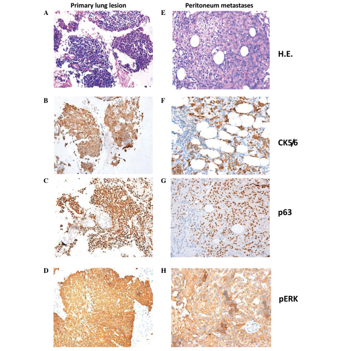
Figure 3.
Hematoxylin and eosin staining and 3,3′-diaminobenzidine immunohistochemistry of the (A-D) primary lung lesion and (E-H) peritoneal metastasis. (A and E) Hematoxylin and eosin staining. (B and F) Cytokeratin 5/6 (positive in cytoplasm). (C and G) p63 (positive in nucleus). (D and H) pERK (positive in cytoplasm and/or nucleus) (magnification, ×200).

