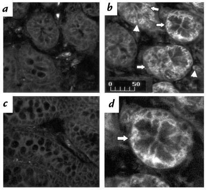
Figure 3.
Increased NTR1 protein expression during toxin A colitis in rats. Rat colonic loops were injected with either toxin A (5 μg) or buffer (control). Animals were sacrificed after 2 h, and colonic tissues were processed for immunohistochemical detection using a rabbit polyclonal antibody directed against the NH2-terminal amino acids 1–28 and 50–69 of the rat NTR1 receptor. Sections were examined by confocal microscopy. (a) Buffer-exposed colon (2 h). (b) Toxin A–exposed colon (2 h). (c) Toxin A–exposed colon (2 h) incubated with the NTR1 antiserum, which was preincubated with an excess of the two NH2-terminal peptides of the NTR1 described above. (d) Higher magnification of b. Note the presence of signal for NTR1 in the colonic mucosa, in particular the colonic epithelial cells (arrows in b and d), and in cells in the lamina propria (arrowhead). Note also the increased immunoreactivity after 2 h exposure to toxin A (b) compared with control (a). Preabsorption of the NTR1 antiserum with an excess of the receptor peptides used to generate the antibody causes complete disappearance of positive staining in toxin A–exposed colon (c). Results are representative of three experiments for each experimental condition. Scale bar: 50 μm.

