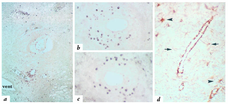
Figure 3.
Expression of CXCR3 and IP-10 in MS lesions. (a) Numerous CXCR3-immunoreactive cells are present in perivascular inflammatory infiltrates in an acute MS lesion in white matter surrounding a ventricle (vent). ×80. (b) CXCR3-immunoreactive cells in a perivascular infiltrate exhibit similar morphology and distribution to CD3-positive T cells (c). ×170. (d) Immunohistochemistry for IP-10 reveals staining of the cell bodies of reactive astrocytes (arrows). Intense staining is also seen in processes, consistent with astrocytic end-feet that extend to, and surround, a blood vessel. Scattered reactive astrocytes in the surrounding parenchyma also express IP-10 (arrowheads). ×300. CXCR3, type 3 CXC chemokine receptor.

