Figure 5.

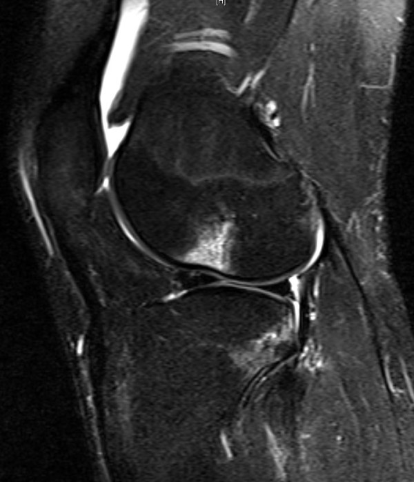
Sagittal fat-suppressed T2-weighted image shows bone contusions in the lateral femoral condyle and posterolateral tibia plateau.

Sagittal fat-suppressed T2-weighted image shows bone contusions in the lateral femoral condyle and posterolateral tibia plateau.