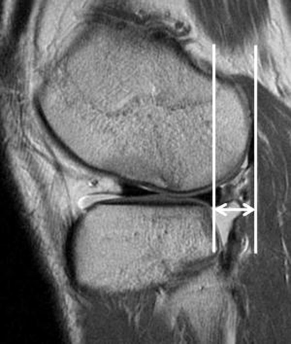
Figure 7.

Anterior translation of the tibia relative to the femur is measured through the middle of the lateral femoral condyle on sagittal images. Two lines are drawn parallel to the cephalocaudal axis of the image: one crossing the posteriormost point of the posterolateral tibia plateau and the other crossing the posteriormost point of the lateral femoral condyle. Anterior translation is determined by the distance in millimeters between these two lines. A translation of ≥7 mm is considered to be a positive indicator of ACL injury.
