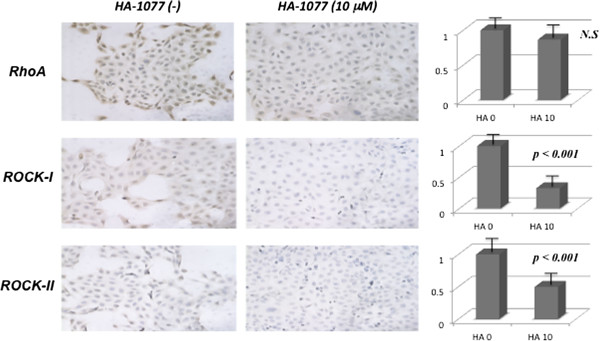
Figure 2.

Immunohistochemical staining using anti-RhoA, anti-ROCK-I and anti-ROCK-II monoclonal antibodies in 5637 (x20 magnification). 70-80% of tumor cells showed moderate to strong cytoplasmic staining reaction for anti-RhoA antibody, and weak to moderate cytoplasmic staining for anti-ROCK-I and anti-ROCK-II antibodies. HA-1077 reduced the staining reactivity for anti-ROCK-I and anti-ROCK-II antibodies to very weak staining in only 20-30% of tumor cells, while it did not decrease the staining for anti-RhoA antibody with weak to moderate staining.
