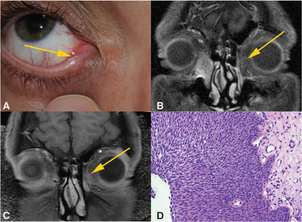
FIG.
A, External photograph showing the lesion emanating from inferior punctum. B, T1 coronal MRI scan showing enhancement of right lacrimal sac with extension into the nasolacrimal duct. Yellow arrow indicating left-sided signal flare suggestive of developing neoplasm. C, T1 coronal MRI scan showing a 7-mm enhancing lesion of the left lacrimal sac. D, High-power hematoxylin and eosin stain of the left lacrimal sac biopsy which reveals proliferation of atypical squamous cells (hematoxylin-eosin).

