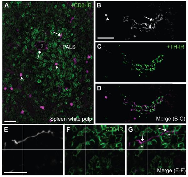
Figure 11.
Identification of neuronal and nonneuronal cholinergic structures in the spleen. A: Fluorescent neuronal fibers along with immune cells were found in the splenic white pulp (20×, 7 optical plans, 1-μm step). B–D: Varicose TH-positive fibers (green) and putative cholinergic fibers frequently intermingled in periarteriolar areas of the white pulp (20×, 8 optical plans, 0.68-μm step). As in other tissues, tdTomato-containing fibers were never TH immunoreactive. E–G: Notably, individual fluorescent fibers were observed around arterioles and occasionally branched into the adjacent periarteriolar lymph sheaths where they contacted CD3-positive cells (green) (40×, 1 optical plan). In E and G, tissue is labeled with an anti-DsRed antibody (white or magenta). Asterisks are positioned over lymphocytes in close proximity to a cholinergic fiber (<1 μm or apparent contact in a single plan). Lastly, isolated immune cells populated the white pulp. Arrows indicate fluorescently labeled neuronal fibers, and arrowheads point to representative cell bodies of immune cells. Abbreviations: a, arteriole; PALS, periarteriolar lymph sheath. Scale bar = 20 μm in A; 50 μm in B (applies to B–D); 10 μm in E (applies to E–F).

