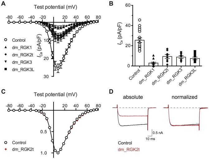
Figure 2. Fruit fly RGK-like protein homologs reduce ICa density in rat sympathetic neurons.
A. I-V plots in which mean ± SEM ICa density is plotted versus test potential. ICa was evoked and acquired as described for Fig. 1. Control neurons (open circles) were not injected with cDNA (n = 17). Neurons previously injected with Drosophila melanogaster RGK protein cDNA clones (50 ng/µl approximately 18–24 hours prior to recording) are depicted with filled symbols: dm_RGK1 (triangle, n = 13); dm_ RGK2t (diamond, n = 8), dm_RGK3 (inverted triangle, n = 8), and dm_RGK3L (square, n = 9). B. Category plot of data shown in panel A for ICa density at +10 mV. Mean ICa density for neurons expressing RGK protein clones differed significantly (P<0.05) from control (one-way ANOVA followed by Dunnett's multiple comparisons test). C. Normalized I-V plots for control (open circle) and dm_RKG2t expressing (red filled diamond) neurons. Data, from panel A, was normalized to the maximal ICa density and plotted to illustrate similarity of voltage-dependence. D. Exemplar ICa traces acquired at +10 mV from control (black) and dm_RGK2t (red) expressing neurons. Traces are depicted without (left) and with normalization (right) to maximal current. Dotted line represents zero current level.

