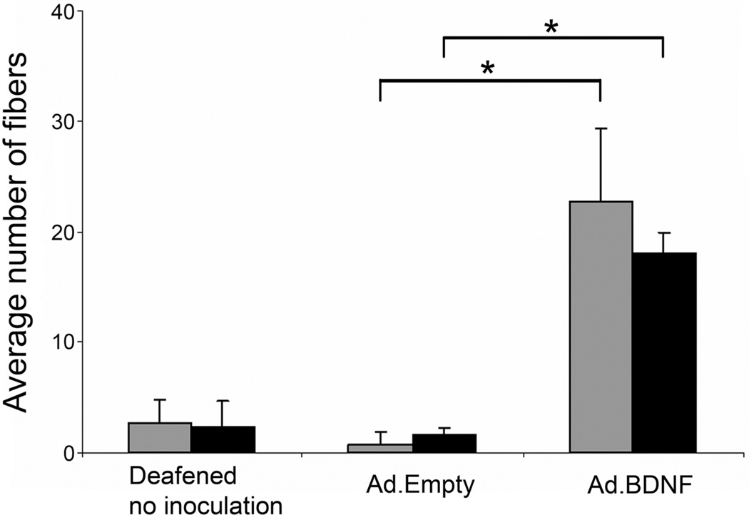
Figure 2.
Quantitative analysis of nerve fibers projecting into the basilar membrane area in deafened ears that have undergone either no treatment, or treatment with Ad.Empty or Ad.BDNF. Deafened animals inoculated with Ad.Empty had similar counts to untreated animals, whereas animals inoculated with Ad.BDNF had a significantly greater average nerve fiber count in the 1st and 2nd turns at 14 (gray bars) and 30 (black bars) days after deafening. (*) indicates p<0.01. Brackets are standard deviation and statistical comparison was done by Student t-test. (Reprinted with permission from Shibata et al., 2010)

