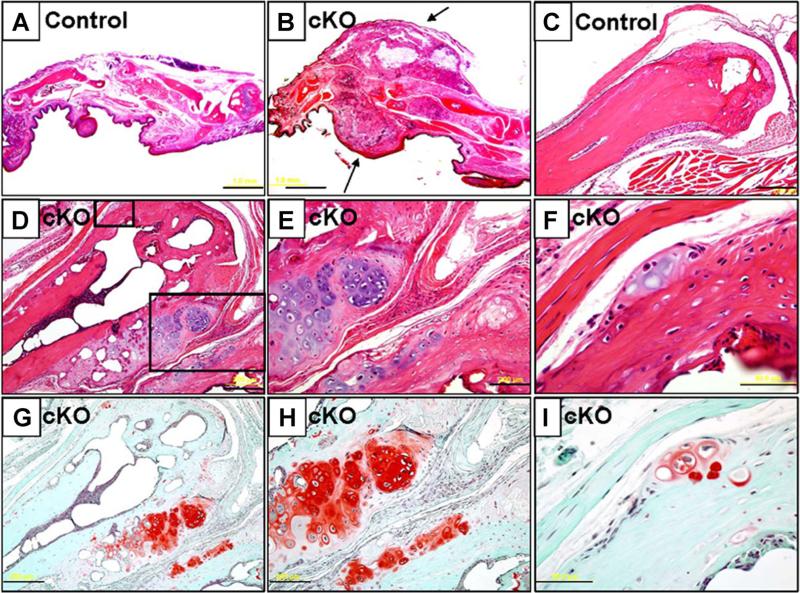
Fig. 4.
Histological assessment of hands of SHP2 cKO mice. (A, B) H&E-stained sagittal sections of digits from control (A) and SHP2 cKO mice (B) at 12 weeks of age. Note an extra bony mass covered by a fibrous cap around the metacarpal-phalangeal joint in the SHP2 cKO mice. (C–F) H&E-stained sections of distal metacarpal bones of control mice (C) and SHP2 cKO mice (D–F) at 12 weeks of age. Extra bony nodules were observed in the distal metaphyseal region of the metacarpal bone of the SHP2 cKO mice. The boxed fields (D) were magnified (E, F). (G–I) Serial sections of distal metacarpal bones of SHP2 cKO mice corresponding to images (D–F) were stained with Safranin O to reveal cartilaginous matrix (red). In particular, note strong staining of excess bony region around the metaphysis of the metacarpal bone. Note that early stage of disease was developed in the periosteum or perichondrium region (F, I). Scale bars = 1 mm (A, B), 200 μm (C, D, E, G, H), 50 μm (F, I).

