Figure 3.
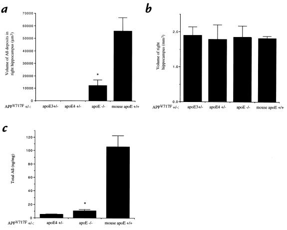
Quantitation of Aβ deposition in APPV717F+/– mice. The total volume of Aβ-IR deposits in the right hippocampus (a) as well as the volume of the right hippocampus itself (b) was determined in each group of APPV717F+/– mice: apo E3+/– line 37 (n = 4), apo E4+/– line 22 (n = 7), apo E–/– (n = 8), and mouse apo E+/+ (n = 8). There was a significantly smaller volume of Aβ-IR deposits (P < 0.05) in the mice expressing human apo E3 compared with mice expressing no apo E or mouse apo E. The volume of the hippocampus was not statistically different between the different groups. Aβ ELISA for total Aβ in the left hippocampus of APPV717F+/– mice that were apo E4+/–, apo E–/–, and mouse apo E+/+ (c) revealed the same pattern of results as was found for Aβ-IR deposits histologically. *P < 0.05 comparing apo E–/– mice with apo E3+/– and apo E4+/– mice.

