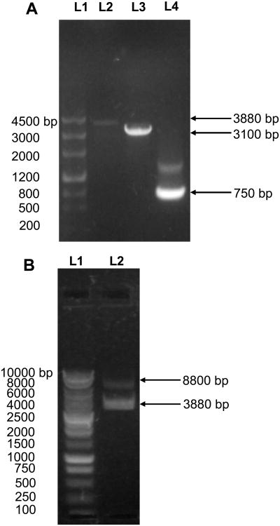
Figure 2.
Characterization of egfp-mexB fusion gene using agarose gel electrophoresis: (A): (L1) DNA markers/ladders in base pair (bp); (L2) The egfp-mexB fusion gene amplified using both egfp P1 and mexB P2 as primers (Figure S1A-B:III); (L3) The ds-mexB gene amplified using the genomic DNA of P. aeruginosa as a template, and mexB P1 and P2 as primers (Figure S1B-I); (L4) The ds-egfp gene amplified using the plasmid (pEGFP) as a template and egfp P1 and P2 as primers. Arrows point to the PCR products of 750, 3100 and 3880 bp, which agree well with the number of base pairs of egfp, mexB and egfp-mexB fusion gene. The sequence of egfp-mexB is characterized using DNA sequencer and shown in Figure S2 in on-line supplementary information. (B): (L1) DNA markers/ladders in bp; (L2) The pMMB67EH-EGFP-MexB vector digested by a pair of restriction enzymes, SalI/HindIII. Arrows point to the digested vector plasmids at 8800 bp and egfp-mexB fusion gene at 3880 bp.

