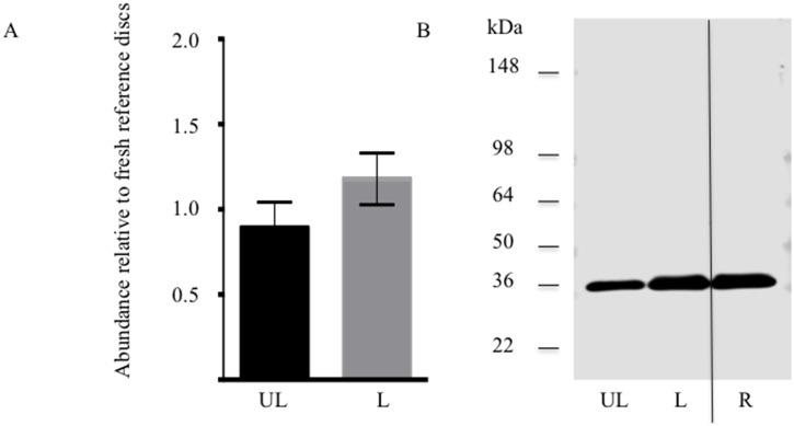
Figure 4. Extractable chondroadherin in loaded and unloaded discs.
NP tissue of loaded (L) and unloaded (UL) IVDs were analysed by western blotting after 14 days of culture. The IVDs were injected 100 µg trypsin prior to loading. A, Semi-quantitative analysis of chondroadherin with a molecular weight of about 36 kDa. Each sample was evaluated in duplicate on 2 separate western blots. Band intensity was normalized to reference samples (R) included on each blot. B, representative Western blot of pooled extracts. (n 6 discs from different animals). Error bars represent SEM.

