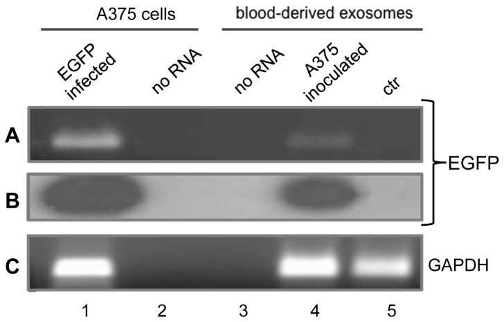
Figure 3. EGFP-specific RNA in circulating blood from A-375/EGFP-inoculated mice.
A: Ethidium bromide staining of specific RT-PCR products from RNA extracted from EGFP-infected A-375 cells (lane 1) and blood-purified extracellular exosomal fraction from inoculated (lane 4) and non-inoculated (lane 5) mice. No RNA and no primers were added to the amplification mix in lanes 2 and 3, respectively. B: EGFP hybridization pattern. The gel in A was blotted on filter, hybridized with 32P-end labelled EGFP-specific probe, washed and autoradiographed. C: GAPDH-specific amplification products from the same samples.

