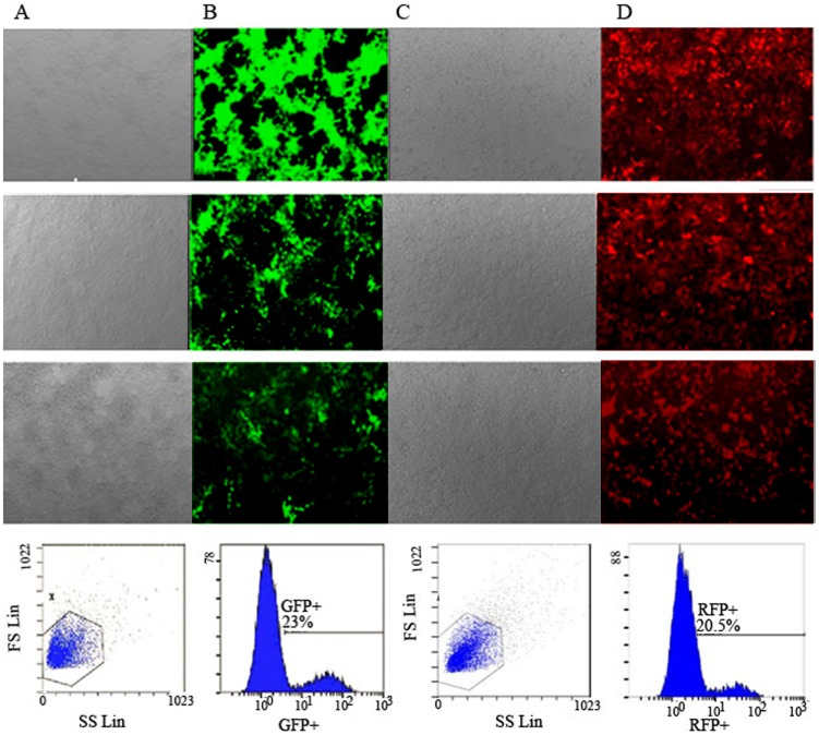
Figure 1. Titers of pLenO-GFP-NS1 and pLenO-RFP-NS2 were assayed by flow cytometry.
A, B: Infection of HEK293T cells with different dilutions of pLenO-GFP-NS1 under light microcopy and fluorescence microscopy respectively (×100); C, D: Infection of HEK293T cells with different dilutions of pLenO-RFP-NS2 under light microcopy and fluorescence microscopy respectively. From above to below represent the concentration of 1.0 ul, 0.1 ul, 0.01 ul and representative images of flow cytometry (0.01 ul).

