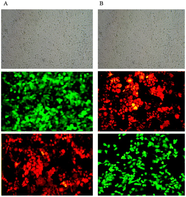
Figure 2. Efficiencies of lentiviral infections to 16 HBE were assayed by fluorescence microscopy and indirect immunofluorescent technology.
A: Infection of 16 HBE by pLenO-GFP-NS1 (MOI = 10); B: Infection of 16 HBE by pLenO-RFP-NS2 (MOI = 10). From above to below represent images under light microcopy, fluorescence microscopy and indirect immunofluorescent assay (×200).

